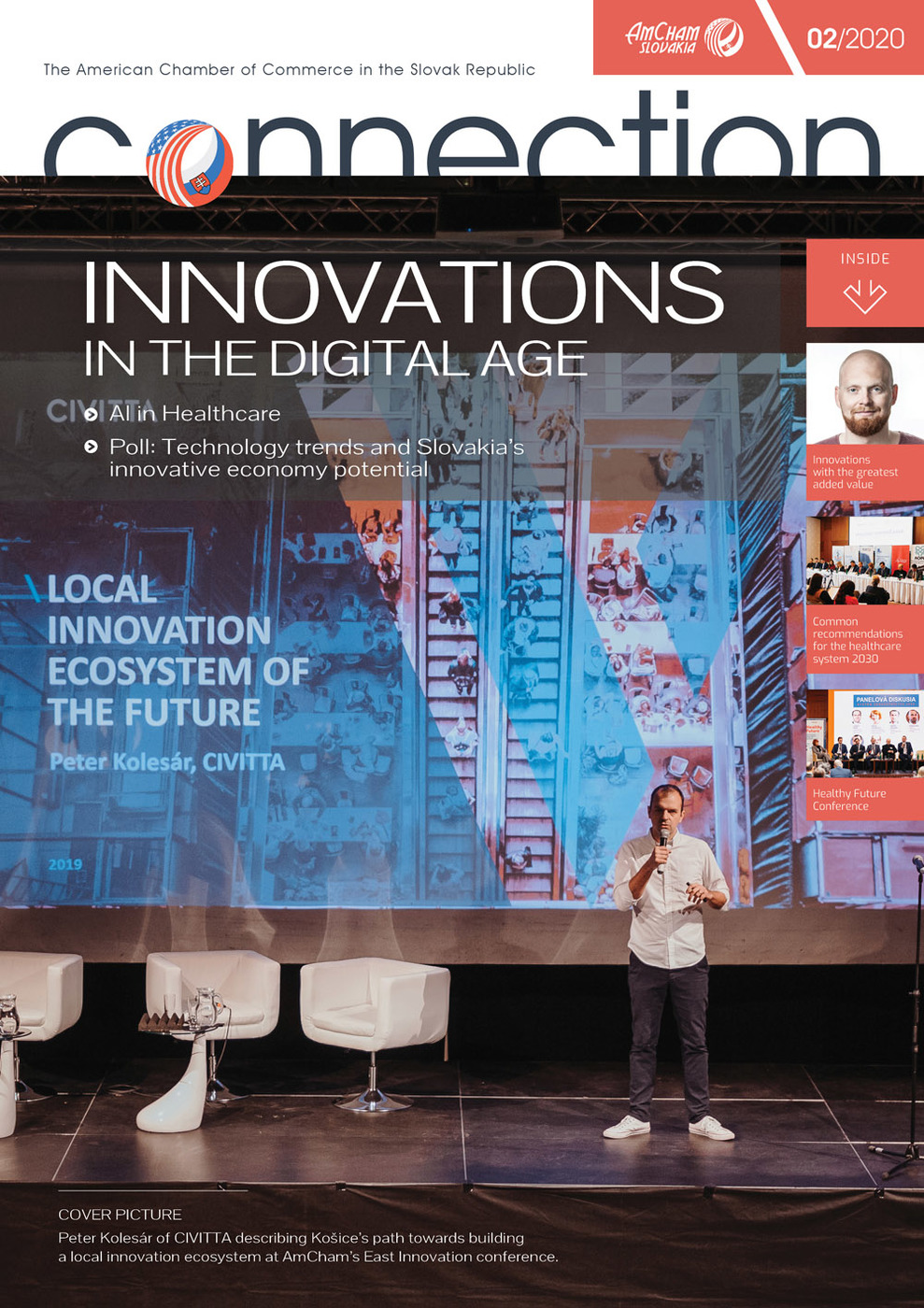
Computer programs use “deep learning” to train themselves to spot diseases by analyzing thousands of medical data - images, or signals. They draw on data from past health records to spot similarities in conditions and make an accurate diagnosis without human assistance. This article discusses some interesting proof-of-concept use cases developed within GlobalLogic in Košice.
Automatic heart attack detection from the ECG paper
Slovakia has one of the highest mortality rates for cardiovascular diseases among the OECD countries. In the case of myocardial infarction (also known as heart attack), the time between the occurrence of infarction and the appropriate medical help needs to be shrunk down as much as possible. Literally, every minute matters.
A typical scenario looks like this - an ambulance is called after a subjective pain in the chest of the patient is reported. The patient is taken to the nearest hospital, where a specialist performs diagnosis (based on ECG), and determines that the patient needs to be quickly transported to a specialized hospital because proper treatment cannot be offered on the spot. But in the end, it may be too late.
Nowadays there are already available solutions on the market, helping to save precious time by the diagnosing the patient’s condition on the spot or during transport. This ECG data is evaluated by specialists (cardiologists, in this case) who are not present in the field, but who receive the data sent by the ambulance rescuer via a mobile application. The prototype we developed aims to improve the existing solution and get the evaluation data results without the need to contact any other person or specialist.
First, the app automatically extracts the ECG leads from the image using a deep convolutional neural network that was trained on manually labeled images. After that, the leads are analyzed by another neural network, analyzing the signal patterns, finally outputting the probability of a heart attack. Subsequently, the ambulance can decide right away whether to bring the patient directly to the specialized hospital where he can undergo the proper treatment.
Knee implant size prediction
Imagine that you have problems with your knee and the specialist recommends replacing it with a knee implant. Since every person has a different size and shape of the knee joint, the question arises how to determine the proper size for your knee. One way how it is performed these days, is to try different standardized sizes directly during the surgery. This solution is definitely not optimal. It would be best to know the proper size before the surgery, so that the preparation can be more effective. The obvious solution is to perform a CT scan of your knee, which directly creates a 3D model of your knee. Based on this model, it is possible to either accurately determine the closest standardized size to your knee or alternatively, to use 3D printing technologies to construct a totally personalized version of your knee joint.
However, a CT scan is not completely harmless, as it exposes you to a non-negligible amount of radiation. Therefore, the idea was to determine the knee implant size from an X-ray image alone, since the radiation doses for X-ray scans are about 100 times smaller than those of CT scans. Similarly, as in the previous project, we have successfully applied a family of deep convolutional neural networks to extract the knee joint from an X-ray image and then to infer the whole 3D model of a knee joint. This way, one can predict the knee implant size with acceptable accuracy while using only one X-ray projection.
Automatic wound measurement
The number of diabetes cases has increased rapidly in recent decades. The combination of reduced blood flow, as well as neuropathy in the feet, increases the chance of foot ulcers that can lead to limb amputation. The healing of ulcers is very demanding and requires daily interaction. To coordinate the treatment, there is a need to record a change in wound morphology. Patients either have to visit a doctor very often or manually measure the wound dimensions using a ruler. This is certainly not pleasant and the measurements may not be accurate.
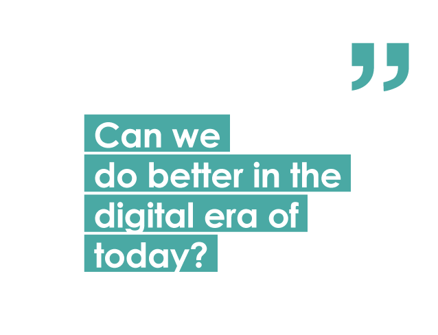 Can we do better in the digital era of today? Are smartphones smart enough to provide the necessary information in a single photo? Phones are already equipped with many sensors, such as depth sensors and cameras. They can create a spatial map of the scanned environment with high accuracy. As a result, in collaboration with machine learning algorithms, we can obtain a 3D ulcer model on an everyday basis and track the progress of treatment. The patient can quickly, accurately and in a contactless way measure the size of the wound in the comfort of his home. Expert’s guidance is also involved as the doctor monitors the data shared by patients through an online portal. Thus, one skilled in the art can easily perform a more detailed analysis of the wound from the 3D model without the patient having to visit the clinic. In practice, telemedicine is no longer just a vision of the future.
AI and doctors fighting diseases alongside
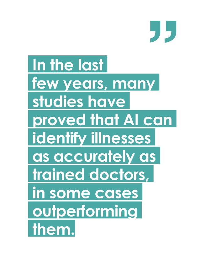
Diagnosis of disease using deep learning algorithms holds enormous potential. In the last few years, many studies have proved that AI can identify illnesses as accurately as trained doctors, in some cases outperforming them.
Innovations in the Digital Age
- Innovations with the greatest added value
- The arrival of artificial intelligence
- AI and doctors fighting diseases alongside
- Common recommendations for the healthcare system 2030
- New space for Slovak businesses
- Software robotization redefining white-collar industry
- The workplace of the future
- Fintech battlegrounds: Europe vs. the U.S.
- Transformative technologies and Slovakia’s path towards an innovative economy
- United States visa options for the Slovak entrepreneur, business or start- up
- The level of R&D matters!
- Science parks as ecosystems for efficient technology transfer and innovation
Follow us